
Thyroid cancer: types, how to recognize it, and how it is treated today
In most cases, thyroid cancer is curable. This article covers diagnosis, the different types, and how it is treated.
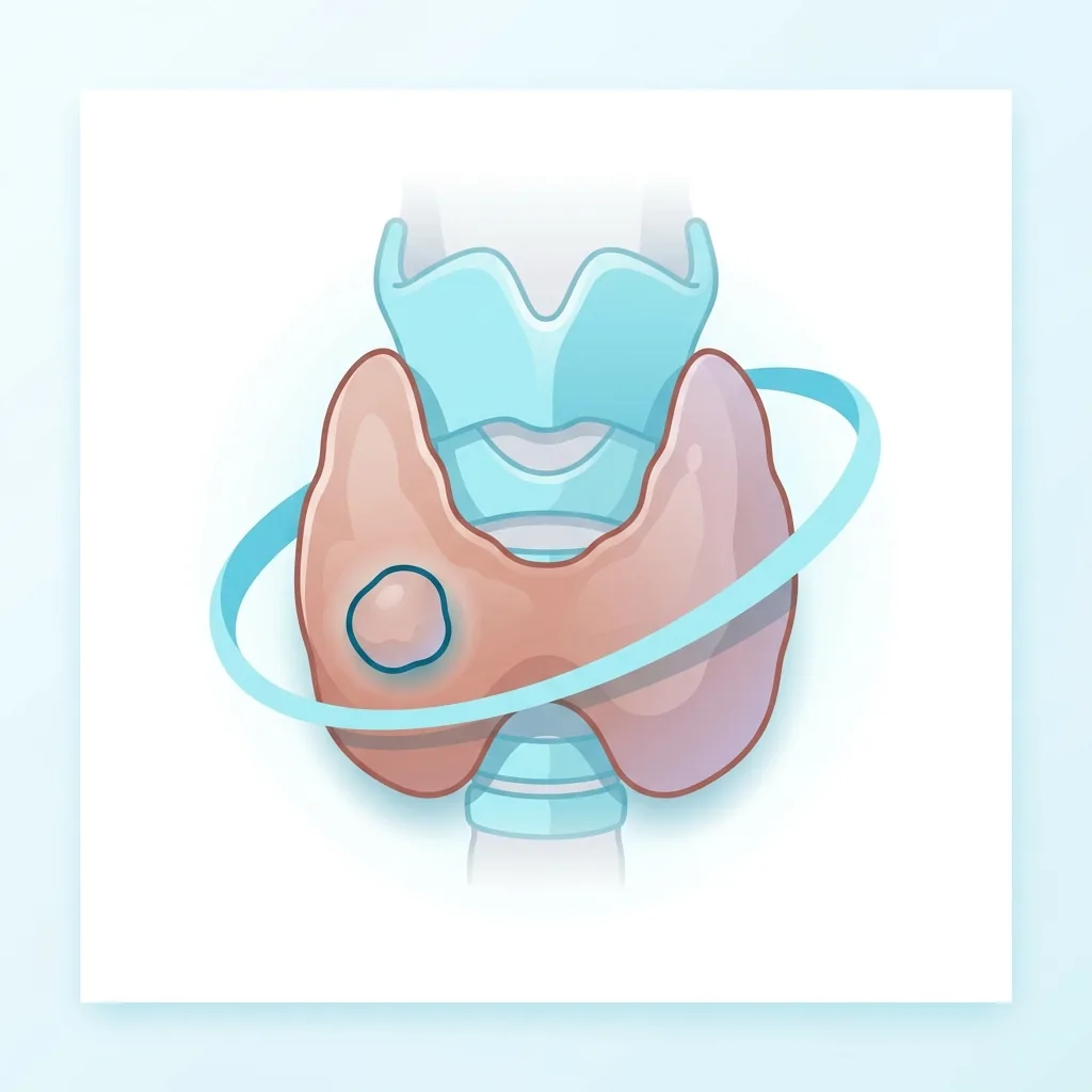
What is thyroid cancer?
Thyroid cancer is the most common endocrine cancer, although it accounts for only about 1% of all cancers. The number of cases has been rising worldwide, mainly because ultrasound can detect small papillary cancers. With modern treatment, most thyroid cancer cases have a very good chance of recovery.
The main types of thyroid cancer
Papillary thyroid cancer (PTC) accounts for 80–85% of cases, grows slowly, often metastasizes to cervical lymph nodes, and 10-year survival in low-risk patients exceeds 95%. Follicular thyroid cancer (FTC) accounts for 10–15% of cases and usually spreads hematogenously to the lungs and bones. Medullary thyroid cancer (MTC) accounts for 2–4% of cases, arises from C cells, can be sporadic or inherited (MEN2 syndromes), and produces calcitonin. Anaplastic thyroid cancer accounts for less than 2% of cases, is aggressive, occurs mainly in the elderly, and has a poor prognosis. Hürthle cell (oncocytic) carcinoma is a subtype of follicular thyroid cancer.
Symptoms
Most patients have a painless nodule in the neck that can be seen on ultrasound or felt on examination. Warning signs are rapid growth, a hard (firm) or fixed consistency, enlarged cervical lymph nodes, hoarseness, and difficulty swallowing or breathing. In medullary thyroid cancer (MTC), diarrhea and flushing may occur. Anaplastic cancer can cause a hard, fixed mass that grows rapidly, sometimes in just a few weeks.
Blood tests
TSH is measured. Calcitonin is tested when medullary thyroid cancer (MTC) is suspected, for example with a family history of MEN2 syndrome, a suspicious ultrasound finding, or an elevated CEA level. Thyroglobulin has no diagnostic value before surgery in differentiated thyroid cancer, but becomes the main tumor marker after total thyroidectomy. Thyroglobulin values should be very low or undetectable. At the same time, anti-thyroglobulin antibodies must be measured, since they can interfere with the thyroglobulin assay. Before surgery, calcium, parathyroid hormone, and vitamin D levels are also assessed; in MTC suspicion, RET gene mutation testing and metanephrines are checked to rule out pheochromocytoma.
Imaging studies
Ultrasound of the thyroid and neck is the first-line examination for detecting a nodule, assessing its risk according to the TIRADS classification, and mapping the lymph nodes. Lymph nodes with suspicious morphology are biopsied. Fine-needle aspiration biopsy allows a cytological diagnosis (according to the Bethesda category). For larger or higher-risk tumors, additional neck and chest computed tomography is used (without iodine contrast if radioactive iodine therapy is planned in the near future), neck MRI to assess retrotracheal or mediastinal spread, and a whole-body radioactive iodine scan or FDG-PET scan to detect distant metastases. When calcitonin levels are very high in suspected medullary thyroid cancer (MTC), a neck ultrasound, chest and abdominal CT, and a bone scan or MRI are performed.
Treatment and treatment options
Surgery is the most important part of treatment. In low-risk unilateral papillary cancer, when the tumor diameter is under 4 cm, removal of a lobe (lobectomy) is usually enough. For higher-risk, multifocal, or larger tumors, a total thyroidectomy is performed. When lymph nodes are involved, central and/or lateral cervical lymph node dissection is added. Postoperative radioactive iodine (I-131) ablation is used for specific intermediate- and high-risk differentiated cancers. Long-term TSH suppression therapy with levothyroxine is applied to keep TSH below 0.1 mIU/L in high-risk patients, between 0.1–0.5 mIU/L in intermediate-risk patients, and between 0.5–2.0 mIU/L in low-risk patients. In anaplastic and advanced medullary thyroid cancer, systemic therapy with tyrosine kinase inhibitors (e.g., selpercatinib, cabozantinib, larotrectinib, dabrafenib/trametinib) may be needed, and radiation therapy is used when necessary.
Follow-up after surgery
After a total thyroidectomy with or without radioactive iodine therapy, TSH, fT3, thyroglobulin (TG), and anti-thyroglobulin antibodies (anti-TG) are typically monitored every 6–12 months. A neck ultrasound is performed at 6–12 months, and every 1–2 years in low-risk disease. A rising thyroglobulin level can be an early sign of disease recurrence. In medullary thyroid cancer (MTC), calcitonin and CEA are monitored initially every 3–6 months and later every 6–12 months. The need for further investigations depends primarily on changes in tumor markers over time. Lifelong follow-up by an endocrinologist is recommended.
Medical disclaimer. This article is for general educational purposes and does not replace consultation, diagnosis or treatment by a qualified healthcare professional. Always discuss your individual situation with your own doctor.