
Thyroid nodules: assessment, TIRADS, and biopsy
Ultrasound studies have shown that 50–60% of adults have thyroid nodules. Most of them are not malignant.
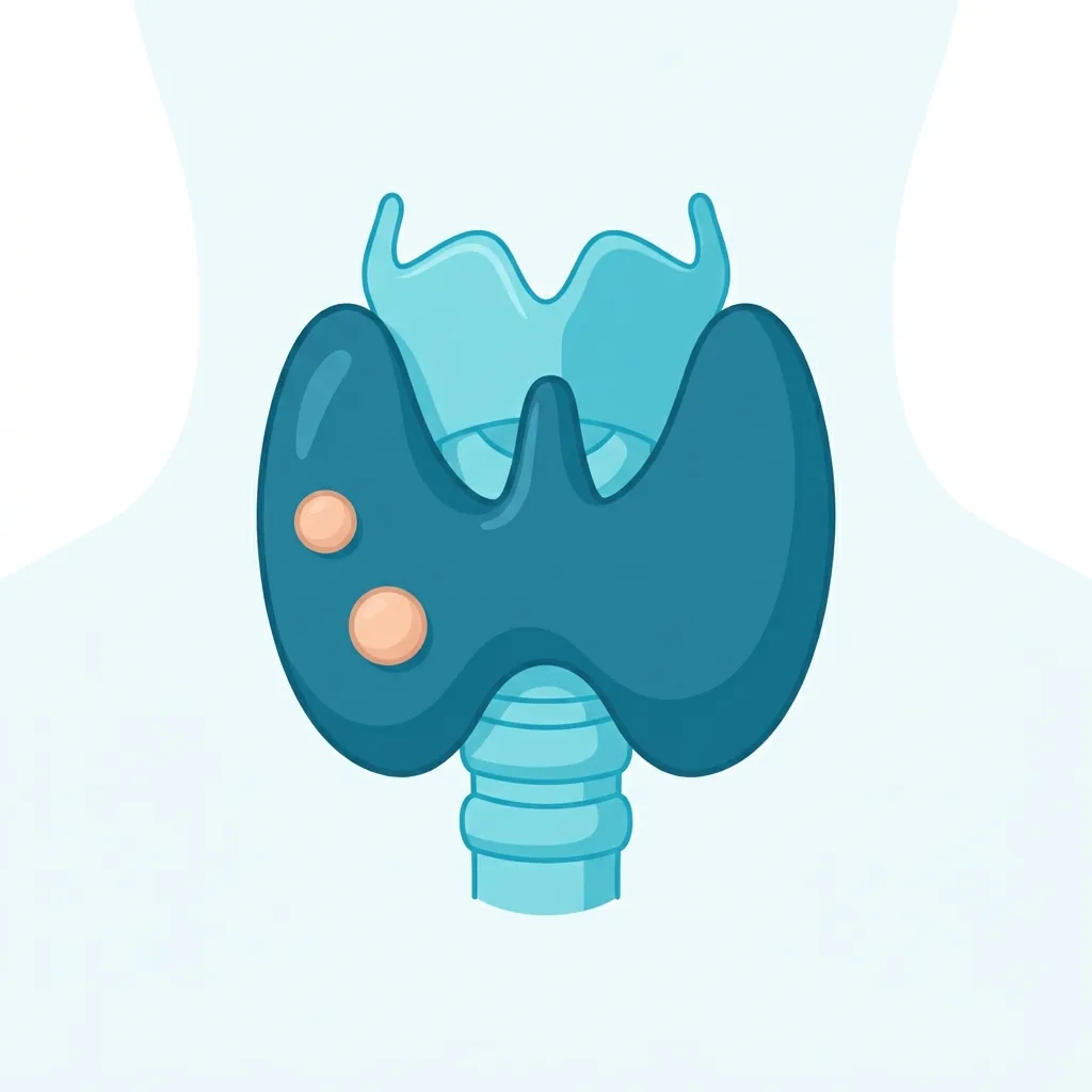
How many people have thyroid nodules?
About 5% of adults have palpable nodules, but ultrasound detects them in 50–60% of cases. About 5–10% of nodules are malignant. The rest are not malignant and are colloid nodules, adenomas, cysts, or pseudo-nodules associated with Hashimoto's disease.
Symptoms
Most nodules cause no symptoms and are found incidentally during another examination. Large nodules can cause a visible or palpable neck swelling, a feeling of pressure, difficulty swallowing, hoarseness, or, in rare cases, difficulty breathing. A rapidly growing or firm, fixed-feeling nodule together with enlarged cervical lymph nodes suggests malignancy. Hormonally active nodules can cause symptoms of an overactive thyroid.
Blood tests
In every patient with thyroid nodules, the TSH level should be checked. A TSH below the normal range suggests an autonomously functioning (hot) nodule. Further assessment is then done with scintigraphy, not biopsy, because hot nodules are usually benign. Calcitonin is measured selectively in patients with a family history of medullary thyroid cancer or MEN2 syndrome, as well as when the nodule has suspicious features. A value above 100 pg/ml suggests the possibility of medullary thyroid cancer. If Hashimoto's thyroiditis is suspected, measuring anti-TPO is helpful.
Imaging studies — ultrasound and TIRADS
A thyroid ultrasound is required. Each nodule has a unique combination of cystic, spongiform, mixed, and solid components and its own echogenicity (anechoic, hyperechoic, isoechoic, hypoechoic, or markedly hypoechoic), shape (wider-than-tall vs. taller-than-wide), margins (smooth, ill-defined, lobulated, or with extrathyroidal extension), and echogenic foci (none, "comet-tail" artifacts, macrocalcifications, peripheral rim, or punctate microcalcifications). The combined assessment of all these features determines the TIRADS or ACR-TIRADS category (1–5). The higher the category, the higher the risk of malignancy. Cervical lymph nodes are also evaluated. Scintigraphy is performed only when the TSH level is low.
When to perform a fine-needle aspiration biopsy (FNA)?
For TIRADS 5 nodules that are 10 mm or larger, and TIRADS 4 nodules that are 15 mm or larger, an FNA is usually recommended. According to the ACR-TIRADS system, biopsy is taken from TR5 nodules above 10 mm, TR4 nodules that are 15 mm or larger, and TR3 nodules that are 25 mm or larger. Any suspicious-looking lymph node should also be biopsied. The Bethesda classification is used to evaluate results: I — non-diagnostic, II — benign, III — AUS/FLUS (atypia/follicular lesion of undetermined significance), IV — follicular neoplasm, V — suspicious for malignancy, and VI — malignant. Molecular tests (e.g., ThyroSeq, Afirma) can improve diagnostic accuracy in Bethesda III–IV cases.
Treatment and monitoring
Benign nodules (Bethesda II) usually do not require any treatment. Small benign nodules (20 mm) should be rechecked with ultrasound after 12–24 months and larger or more complex ones every 6–12 months. For cystic nodules, treatment options include ethanol ablation, radiofrequency ablation, or surgery, especially when the nodules grow or enlarge. For "hot" nodules and hyperthyroidism, treatment is either radioactive iodine or surgery. All Bethesda IV–VI nodules are referred for surgical treatment, which can be either removal of a thyroid lobe (lobectomy) or removal of the entire thyroid gland (thyroidectomy). Depending on the risk assessment, the operation may or may not include removal of the cervical lymph nodes (central neck dissection).
How often to come in for follow-up?
Stable benign nodules are usually monitored with ultrasound every 1–2 years, and if they remain unchanged for 3–5 years, the frequency of check-ups can be reduced even further. After a benign fine-needle aspiration biopsy (FNA), if the ultrasound finding is of low suspicion, a repeat ultrasound is performed in 24 months. However, if a benign FNA finding is associated with an intermediate- or high-suspicion ultrasound finding, the ultrasound is repeated in 12 months, and a repeat biopsy is considered when needed.
Medical disclaimer. This article is for general educational purposes and does not replace consultation, diagnosis or treatment by a qualified healthcare professional. Always discuss your individual situation with your own doctor.